Tomography with neutrons and X-rays shows where particles are deposited
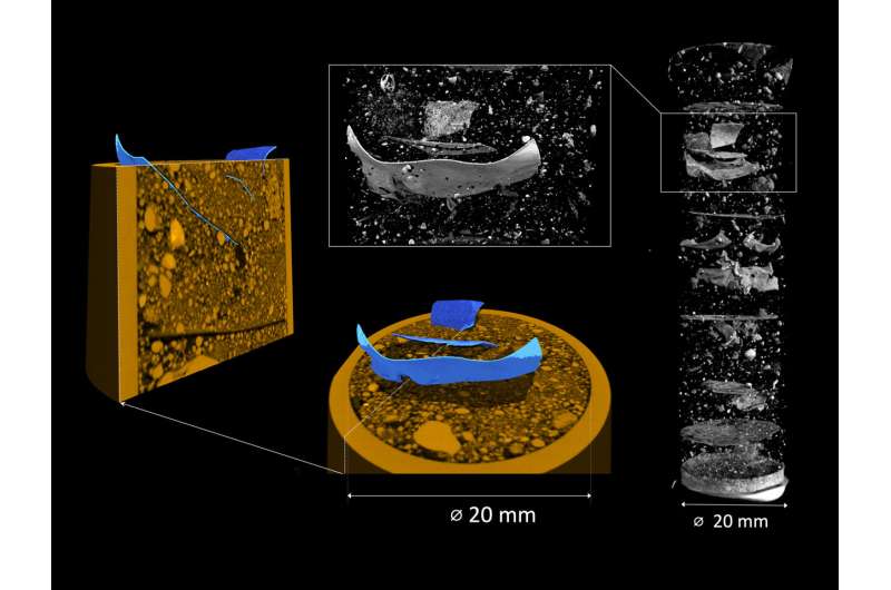
It is a real problem: Microplastic particles are everywhere. Now a team from the University of Potsdam and HZB has developed a method that allows it for the first time to precisely localize microplastic particles in the soil. The 3D tomographies show where the particles are deposited and how structures in the soil are changed.
The method was validated on prepared samples. The team used a special instrument at the neutron source of the Institut Laue-Langevin in Grenoble to carry out neutron and X-ray analyses simultaneously.
Microplastic particles are a major environmental pollutant today. Road traffic accounts for a particularly large proportion: in Germany alone, tire wear is said to generate around one hundred thousand tonnes of microplastics every year, in addition to particles from astroturf, cosmetics, washing powders, clothing, disposable masks, plastic bags and other waste that end up in nature.
Microplastic particles can now be found everywhere. But what happens to these particles in different soils? Do they break up into smaller and smaller pieces and how are they relocated and transported, changing the structures in the soil?
Some of these questions are already being analyzed: A soil sample is floated in a heavy salt solution, whereupon the individual components separate according to density: Plastic and organic particles float to the top, while mineral particles sink.
The mixture of organic material and plastic particles is then treated with hydrogen peroxide, for example, whereby the organic components decompose and the microplastic particles should remain. Although this method makes it possible to determine the quantity and type of microplastic in a soil sample, information is lost as to where exactly these particles accumulate in the soil and whether they change any structures in the soil.
3D tomography with neutrons and X-rays
In their new study, Prof Sascha Oswald (University of Potsdam) and Dr. Christian Tötzke (University of Potsdam and HZB) have now presented a method to answer these questions.
They worked closely with the team led by Dr. Nikolay Kardjilov, HZB, whose expertise went into setting up a unique instrument at the Institut Laue-Langevin, Grenoble: there, samples can be analyzed with neutrons and X-rays to create 3D tomographies simultaneously, i.e. without altering the sample. While neutrons visualize organic and synthetic particles, X-ray tomography shows the mineral particles and the structure they form.
To test the method, Tötzke prepared a series of soil samples from sand, organic components such as peat or charcoal and artificial microplastic particles. In a further series of measurements, he investigated how the roots of fast-growing lupins penetrate the soil samples and how they react to the presence of microplastics.
In the neutron tomograms, the microplastic particles are clearly identified, as can some of the organic components. X-ray tomography, on the other hand, provides an insight into the arrangement of the sand grains, whereas the organic and plastic particles are shown as diffuse voids. When superimposed, a complete image of the soil sample is obtained. This allows the scientists to estimate the size and shape of the microplastic particles, as well as the changes to the soil structure caused by the embedded microplastics.
“This method is quite complex, but it makes it possible for the first time to investigate where microplastic particles are deposited and how they change the soil and its structure,” explains Tötzke. He also analyzed sandy soil from a field near Beelitz, a typical asparagus-growing area in Brandenburg near Berlin, into which he mixed pieces of so called mulch film, a very thin plastic film used to protect the plants.
In “real life” farming it is usually not possible to remove this film after use completely. Remaining film residues are then carried into deeper soil layers during plowing.
“We were able to show that fragments of such films can change the water flow in the soil. Microplastic fibers, on the other hand, created small cracks in the soil matrix,” says Tötzke. It is not yet possible to predict how this will affect the soil’s hydraulic properties, for example its ability to store water. “As droughts and heavy rainfall become more likely with climate change, it is urgent to answer these questions. We now need to investigate this systematically,” says Tötzke.
The work is published in the journal Science of The Total Environment.
More information:
Christian Tötzke et al, Non-invasive 3D analysis of microplastic particles in sandy soil—Exploring feasible options and capabilities, Science of The Total Environment (2023). DOI: 10.1016/j.scitotenv.2023.167927
Citation:
Microplastics in soil: Tomography with neutrons and X-rays shows where particles are deposited (2023, November 16)
retrieved 16 November 2023
from https://phys.org/news/2023-11-microplastics-soil-tomography-neutrons-x-rays.html
This document is subject to copyright. Apart from any fair dealing for the purpose of private study or research, no
part may be reproduced without the written permission. The content is provided for information purposes only.
