
Why Do Some Joint Deformities Hurt While Others Don’t?
Advanced imaging techniques help differentiate symptomatic from asymptomatic discoid lateral meniscus (DLM) cases, with symptomatic ones showing greater meniscal coverage and height. The research aids in surgical planning and sheds light on why symptoms occur in some DLM patients.
Explaining why certain individuals with specific joint deformities experience symptoms, whereas others do not.
With the array of advanced imaging technologies at their disposal, healthcare professionals can diagnose tissue and joint deformities through non-invasive methods with exceptional precision. Yet, a vexing question persists: why do certain patients with specific joint deformities experience symptoms, while others do not?
The meniscus is a piece of cartilage that cushions the knee joint between the femur (thigh bone) and the tibia (shin bone). Some people are born with a congenital morphological variation in their meniscus, called a discoid lateral meniscus (DLM), where the meniscus is thickened on the lateral, or outer side of the knee. DLM malformations cause the lateral meniscus to form a circle rather than a crescent shape, thickening the cartilage and making it more prone to tears. Some patients develop symptoms such as knee pain and a locking, leading to surgery.
Research on Symptomatic vs. Asymptomatic DLM Cases
In order to better understand what factors separate symptomatic DLM cases from asymptomatic cases, a multi-institutional team of researchers led by Dr. Kazuya Nishino of the Graduate School of Medicine at Osaka Metropolitan University analyzed 61 knees with discoid lateral meniscus surgery without dislocation (symptomatic group) and 35 without symptoms but with discoid lateral meniscus detected on MRI (asymptomatic group). The percentage of meniscus was calculated in the coronal and sagittal sections, respectively. The researchers also measured the height of the thinnest and thickest part of the meniscus.
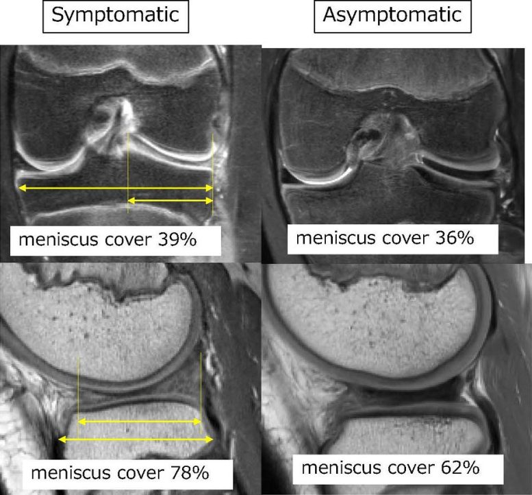
Results showed that coronal and sagittal occupancy rates were higher in the symptomatic group than in the asymptomatic group. Credit: Kazuya Nishino, Osaka Metropolitan University
The results revealed that the percentage of the meniscus covering the tibia in the coronal and sagittal planes was higher in the symptomatic group than in the asymptomatic group. Additionally, the results showed that the meniscal height was greater in the symptomatic group than in the asymptomatic group.
Overall, preoperative imaging is helpful in determining the amount of tissue resection, or removal, required for patients with symptomatic DLM. “Making surgical decisions and plans based on these characteristics is expected to assist in medical treatment. In the future, we will investigate the morphological changes of the meniscus before and after surgery in three dimensions,” said Dr. Nishino.
While the difference in the morphologies of symptomatic vs. asymptomatic DLMs was the most important finding in the study, the MRIs of symptomatic patients also showed meniscal tears or other evidence of instability. The researchers suggest that these morphological features could be responsible for DLM symptoms in symptomatic patients.
Reference: “Comparative analysis of discoid lateral meniscus size: a distinction between symptomatic and asymptomatic cases” by Kazuya Nishino, Yusuke Hashimoto, Takuya Kinoshita, Ken Iida, Shuko Tsumoto and Hiroaki Nakamura, 07 November 2023, Knee Surgery, Sports Traumatology, Arthroscopy.
DOI: 10.1007/s00167-023-07650-2
Advanced imaging techniques help differentiate symptomatic from asymptomatic discoid lateral meniscus (DLM) cases, with symptomatic ones showing greater meniscal coverage and height. The research aids in surgical planning and sheds light on why symptoms occur in some DLM patients.
Explaining why certain individuals with specific joint deformities experience symptoms, whereas others do not.
With the array of advanced imaging technologies at their disposal, healthcare professionals can diagnose tissue and joint deformities through non-invasive methods with exceptional precision. Yet, a vexing question persists: why do certain patients with specific joint deformities experience symptoms, while others do not?
The meniscus is a piece of cartilage that cushions the knee joint between the femur (thigh bone) and the tibia (shin bone). Some people are born with a congenital morphological variation in their meniscus, called a discoid lateral meniscus (DLM), where the meniscus is thickened on the lateral, or outer side of the knee. DLM malformations cause the lateral meniscus to form a circle rather than a crescent shape, thickening the cartilage and making it more prone to tears. Some patients develop symptoms such as knee pain and a locking, leading to surgery.
Research on Symptomatic vs. Asymptomatic DLM Cases
In order to better understand what factors separate symptomatic DLM cases from asymptomatic cases, a multi-institutional team of researchers led by Dr. Kazuya Nishino of the Graduate School of Medicine at Osaka Metropolitan University analyzed 61 knees with discoid lateral meniscus surgery without dislocation (symptomatic group) and 35 without symptoms but with discoid lateral meniscus detected on MRI (asymptomatic group). The percentage of meniscus was calculated in the coronal and sagittal sections, respectively. The researchers also measured the height of the thinnest and thickest part of the meniscus.
Results showed that coronal and sagittal occupancy rates were higher in the symptomatic group than in the asymptomatic group. Credit: Kazuya Nishino, Osaka Metropolitan University
The results revealed that the percentage of the meniscus covering the tibia in the coronal and sagittal planes was higher in the symptomatic group than in the asymptomatic group. Additionally, the results showed that the meniscal height was greater in the symptomatic group than in the asymptomatic group.
Overall, preoperative imaging is helpful in determining the amount of tissue resection, or removal, required for patients with symptomatic DLM. “Making surgical decisions and plans based on these characteristics is expected to assist in medical treatment. In the future, we will investigate the morphological changes of the meniscus before and after surgery in three dimensions,” said Dr. Nishino.
While the difference in the morphologies of symptomatic vs. asymptomatic DLMs was the most important finding in the study, the MRIs of symptomatic patients also showed meniscal tears or other evidence of instability. The researchers suggest that these morphological features could be responsible for DLM symptoms in symptomatic patients.
Reference: “Comparative analysis of discoid lateral meniscus size: a distinction between symptomatic and asymptomatic cases” by Kazuya Nishino, Yusuke Hashimoto, Takuya Kinoshita, Ken Iida, Shuko Tsumoto and Hiroaki Nakamura, 07 November 2023, Knee Surgery, Sports Traumatology, Arthroscopy.
DOI: 10.1007/s00167-023-07650-2
Denial of responsibility! Techno Blender is an automatic aggregator of the all world’s media. In each content, the hyperlink to the primary source is specified. All trademarks belong to their rightful owners, all materials to their authors. If you are the owner of the content and do not want us to publish your materials, please contact us by email – [email protected]. The content will be deleted within 24 hours.