Scans that are 64 million times clearer give a new look at the brain
Fifty years on from American chemist Pal Laterbur detailing the first magnetic resonance imaging (MRI), scientists have marked this historic medical anniversary with the sharpest-ever scans of a mouse brain.
Nearly 40 decades in the making, researchers from Duke University’s Center for In Vivo Microscopy, along with scientists from the University of Tennessee Health Science Center, University of Pennsylvania, University of Pittsburgh and Indiana University, have produced MRI visuals 64 million times sharper than current technology offers.
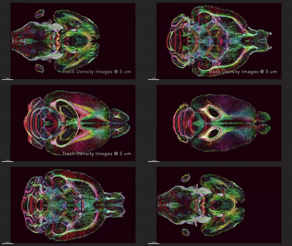
This MRI was able to capture images in such detail that each voxel – the 3D version of a pixel – measured just 5 microns, or five-thousandth of a millimeter. What this means is that while current MRI technology is advanced enough to spot a brain tumor, for example, this sort of clear picture can take things a step further and display organization and far more detailed connectivity.
The MRI was able to capture incredible images of circuitry data throughout the mouse brain, as shown in the video below.
Duke Center for In Vivo Microscopy
The researchers believe that this level of detailed imaging will enable greater understanding of how brains change with age, diet and neurodegenerative diseases such as Alzheimer’s.
“It is something that is truly enabling,” said lead author G. Allan Johnson, professor of radiology, physics and biomedical engineering at Duke. “We can start looking at neurodegenerative diseases in an entirely different way.”
The culmination of almost 40 years of work at the Center for In Vivo Microscopy, this MRI resolution was only made possible with some impressive technology. The team used a powerful 9.4-Tesla magnet (clinical MRIs generally have a 1.5-to-3-Tesla magnet), a set of gradient coils 100 times stronger than in standard scans, and a super-computer equivalent to 800 laptops, all working to capture the single mouse brain.
What’s more, after the MRI visuals were complete, the researchers had the brain tissue scanned by light sheet microscopy. This enabled the scientists to label specific groups of cells, allowing them to watch how neurodegenerative diseases progress over time.
Using sets of mice of varying ages and genetic makeups, the scientists were able to see how the animal’s brain-wide connectivity changed over time and how certain regions, like the memory-related subiculum, changed significantly more than other areas. The images were also able to capture how Alzheimer’s disease breaks down neural networks.
The research paves the way for further technological development to capture the human brain in such detail, which would provide a greater understanding of how tissue changes with age and what interventions could be helpful to stave off degeneration.
“Research supported by the National Institute of Aging uncovered that modest dietary and drug interventions can lead to animals living 25% longer,” Johnson said. “So, the question is, is their brain still intact during this extended lifespan? Could they still do crossword puzzles? Are they going to be able to do Sudoku even though they’re living 25% longer? And we have the capacity now to look at it. And as we do so, we can translate that directly into the human condition.”
The research was published in the journal PNAS.
Source: Duke University
Fifty years on from American chemist Pal Laterbur detailing the first magnetic resonance imaging (MRI), scientists have marked this historic medical anniversary with the sharpest-ever scans of a mouse brain.
Nearly 40 decades in the making, researchers from Duke University’s Center for In Vivo Microscopy, along with scientists from the University of Tennessee Health Science Center, University of Pennsylvania, University of Pittsburgh and Indiana University, have produced MRI visuals 64 million times sharper than current technology offers.
This MRI was able to capture images in such detail that each voxel – the 3D version of a pixel – measured just 5 microns, or five-thousandth of a millimeter. What this means is that while current MRI technology is advanced enough to spot a brain tumor, for example, this sort of clear picture can take things a step further and display organization and far more detailed connectivity.
The MRI was able to capture incredible images of circuitry data throughout the mouse brain, as shown in the video below.

Duke Center for In Vivo Microscopy
The researchers believe that this level of detailed imaging will enable greater understanding of how brains change with age, diet and neurodegenerative diseases such as Alzheimer’s.
“It is something that is truly enabling,” said lead author G. Allan Johnson, professor of radiology, physics and biomedical engineering at Duke. “We can start looking at neurodegenerative diseases in an entirely different way.”
The culmination of almost 40 years of work at the Center for In Vivo Microscopy, this MRI resolution was only made possible with some impressive technology. The team used a powerful 9.4-Tesla magnet (clinical MRIs generally have a 1.5-to-3-Tesla magnet), a set of gradient coils 100 times stronger than in standard scans, and a super-computer equivalent to 800 laptops, all working to capture the single mouse brain.
What’s more, after the MRI visuals were complete, the researchers had the brain tissue scanned by light sheet microscopy. This enabled the scientists to label specific groups of cells, allowing them to watch how neurodegenerative diseases progress over time.
Using sets of mice of varying ages and genetic makeups, the scientists were able to see how the animal’s brain-wide connectivity changed over time and how certain regions, like the memory-related subiculum, changed significantly more than other areas. The images were also able to capture how Alzheimer’s disease breaks down neural networks.
The research paves the way for further technological development to capture the human brain in such detail, which would provide a greater understanding of how tissue changes with age and what interventions could be helpful to stave off degeneration.
“Research supported by the National Institute of Aging uncovered that modest dietary and drug interventions can lead to animals living 25% longer,” Johnson said. “So, the question is, is their brain still intact during this extended lifespan? Could they still do crossword puzzles? Are they going to be able to do Sudoku even though they’re living 25% longer? And we have the capacity now to look at it. And as we do so, we can translate that directly into the human condition.”
The research was published in the journal PNAS.
Source: Duke University
